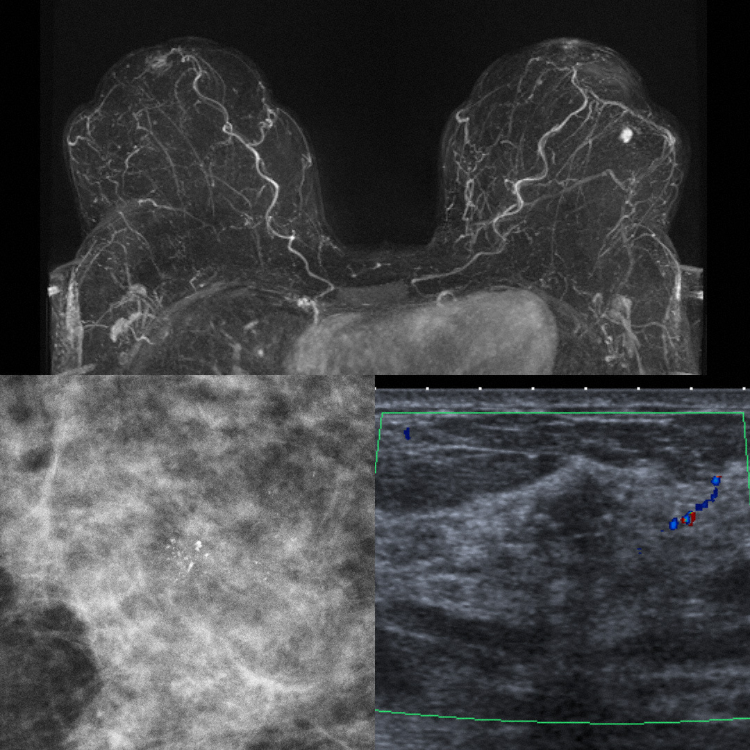
39-year-old asymptomatic female participating in the high-risk multimodality screening program. In the fourth annual screening round with MRI a new enhancing suspicious mass in the left breast was found on MRI (top). Mammography (lower left) showed grouped suspicious calcifications, however the mass itself was not visible on mammography. Tailored second-look ultrasound was able to show a small irregular hypoechoic mass and an ultrasound-guided core biopsy was performed, which demonstrated a Her2-positive invasive carcinoma of no special type with associated ductal carcinoma in-situ (DCIS).
Categories
Case0001
